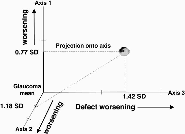
Figure 3.

One field from a series of fields from a patient in this study. Each field in the series is projected independently to each of the six axes of the original vB-ICA-mm space. Only three axes are shown here for clarity. In this depiction, this particular field would be most strongly associated with axis 3 at this point in the original vB-ICA-mm space.
