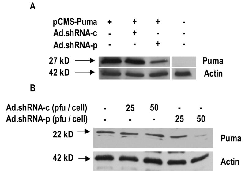
Fig. 2.

Ad.shRNA-p attenuates both exogenous and endogenous Puma expression. A, validation of shRNA by transient transfection. MCF7 cells plated at a density of 1.5 × 10⁶ cells were infected with either the pSuppressorAdeno (non-recombinant virus), Ad.shRNA-c, or Ad.shRNA-p and incubated for 48 h. Cells were then transfected with pCMS-Puma and incubated for an additional 24 h - a total of 72 h. Immunoblot analysis was performed using anti-Puma antibody. The position of exogenous FLAG-tagged Puma band is indicated by an arrow. Actin is shown as a loading control. Results are representative of three independent experiments. B, validation of shRNA in cardiac myocytes. Cells were grown in tissue culture dishes and infected with either Ad.shRNA-c or Ad.shRNA-p for 72 h. Immunoblot analysis was performed using anti-Puma antibody. Actin is shown as a loading control. Results are representative of three independent experiments.
