
Fig. 4.
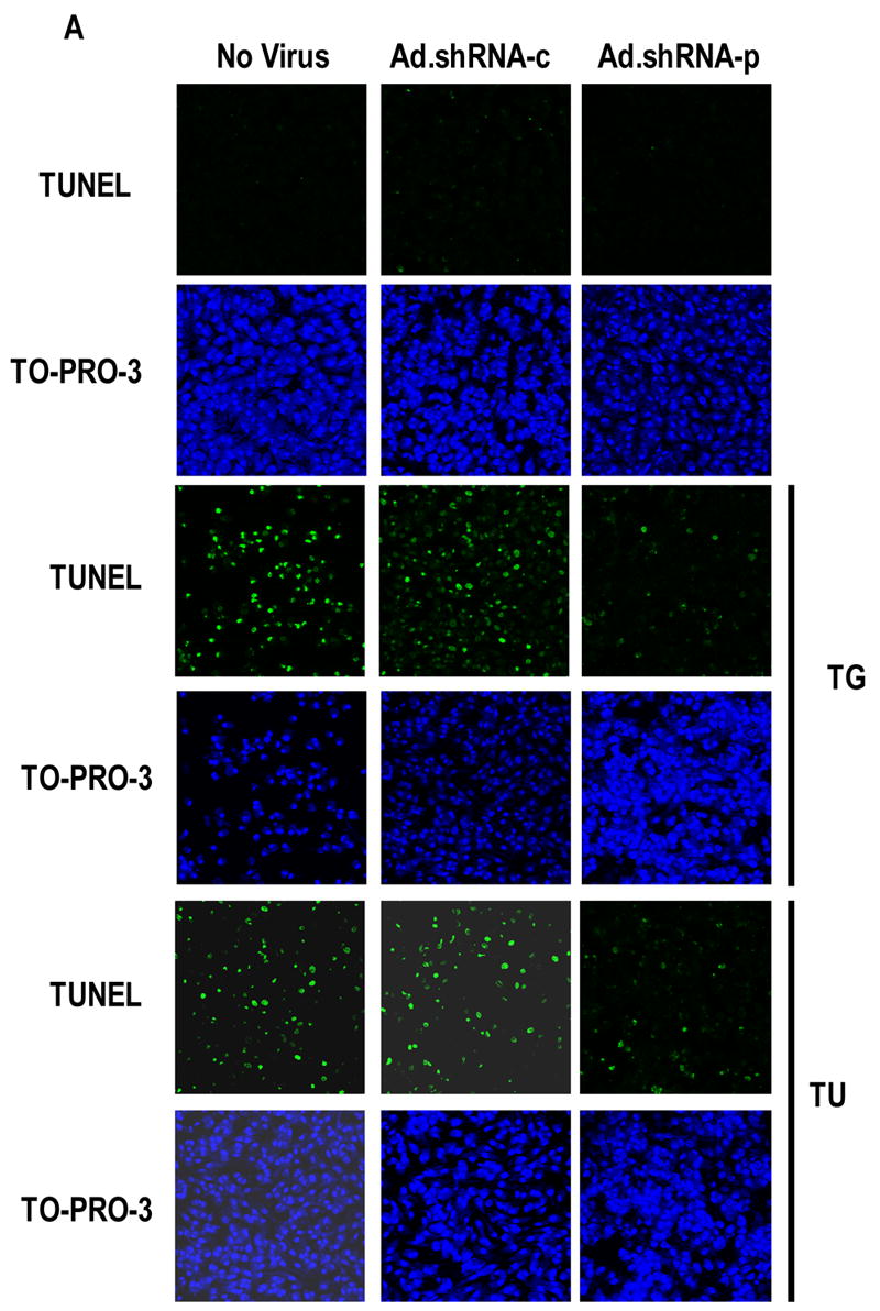
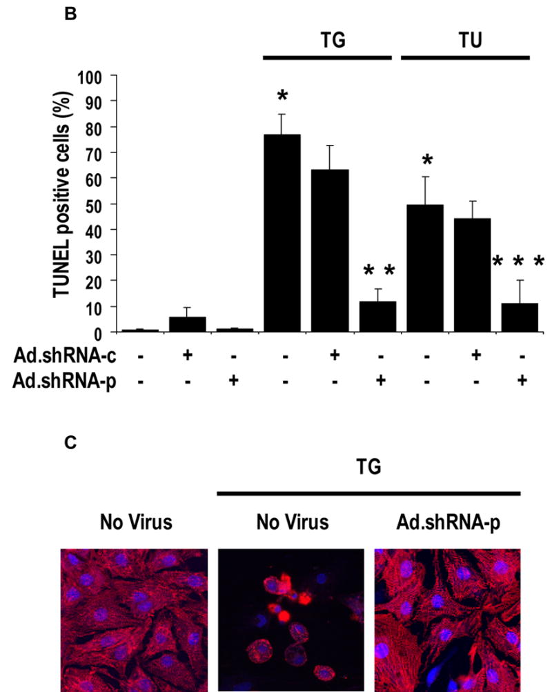
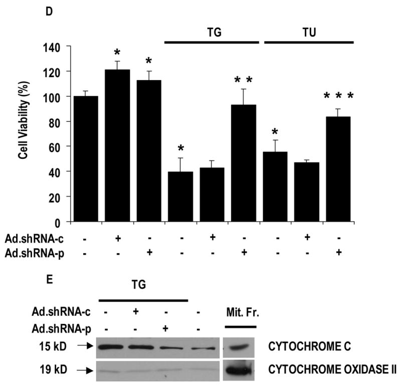
Suppression of Puma attenuates thapsigargin and tunicamycin-induced apoptosis in cardiac myocytes. A, Ad.shRNA-p reduces thapsigargin and tunicamycin-induced apoptosis. Cardiomyocytes were grown in tissue culture dishes and infected with either Ad.shRNA-c or Ad.shRNA-p for 48 h. Cells were then exposed to 3 μM thapsigargin or 100 ng mL−1 tunicamycin for an additional 48 h. Cells were stained for apoptosis by TUNEL assay (green) and nuclei were identified by staining with TO-PRO-3 (blue). Images were taken using a confocal fluorescence microscope at 100X magnification. B, quantification of cardiomyocyte apoptosis. Apoptotic nuclei were scored on the basis of TUNEL positivity as described in A. Data are averaged from three experiments in 10 randomly selected fields (at least 200 cells) from each group. Error bars represent the S.E. of the mean. *, significant difference from the control (p < 0.01). **, significant difference from the TG group (p < 0.01). ***, significant difference from the TU group (p < 0.01). C. High magnification micrographs of cardiomyocytes subjected to treatment with TG, TG plus Ad.shRNA-p, or left untreated, as described in A. Cells were stained with anti-sarcomeric actinin (red) and TO-PRO-3 (blue). Images were taken using a confocal fluorescence microscope at 400X magnification. D, quantification of cardiomyocyte viability. Cardiomyocytes were grown in 96-well tissue culture plates and were infected with either Ad.shRNA-c or Ad.shRNA-p for 48 h. Cells were then exposed to 3 μM thapsigargin or 100 ng mL−1 tunicamycin for an additional 48 h. Cell viability was then assessed using the CellTiter 96 Aqueous One solution. Optical densities were recorded at 490 nm. Data are averaged from 3 independent experiments, each comprising 5 independent replicates. Error bars represent the S.E of the mean. *, significant difference from the control (p < 0.01). **, significant difference from the TG group (p < 0.01). ***, significant difference from the TU group (p < 0.01). E. Ad.shRNA-p prevents thapsigargin-induced cytochrome c release into the cytosol. Cardiomyocytes were grown and treated as described in A. Equal amounts of cytosolic proteins were immunoblotted for cytochrome c and cytochrome oxidase subunit 2 (as control), along with similar amounts of mitochondrial fractions of non-treated cells (Mit.Fr.).