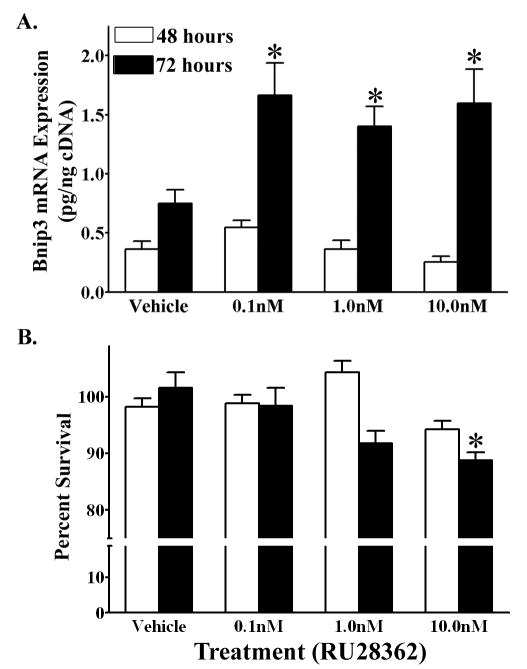
Figure 6.

Glucocorticoids enhance hypoxia induced Bnip3 mRNA expression in primary cortical neurons. (A) Bnip3 mRNA expression (pg/ng cDNA) in primary cortical neurons treated with either RU28362 or vehicle and maintained in hypoxic (1% O2) conditions for 48 (white bars) or 72 (black bars) hours. Each bar represents the mean ± S.E.M. of 9–11 determinations. Data were analyzed by two-way ANOVA (treatment by time). (B) MTT assay to measure percent change in viability of RU28362 or vehicle treated primary cortical neurons maintained in hypoxic conditions for 48 (white bars) or 72 (black bars) hours. The percent survival for hypoxic cultures is relative to vehicle treated hypoxic controls. Each bar represents the mean ± S.E.M. of 12–18 determinations. Data were analyzed by two-way ANOVA (RU28362 by air conditions). Student-Newman Keul’s posthoc test was used to indicate groups with significant differences (*p<0.05) in (A) Bnip3 mRNA levels relative to vehicle treated controls and (B) percent survival relative to hypoxic vehicle- treated cultures.
