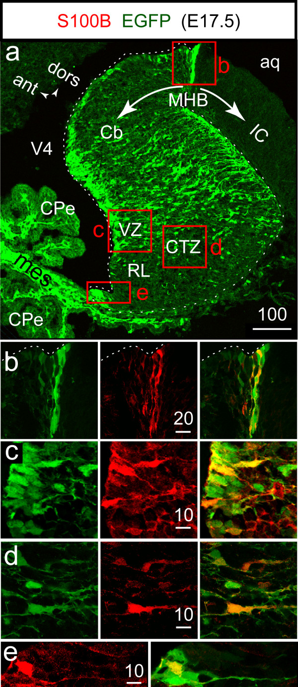
Figure 1.
S100B gene driven expression of EGFP in S100B+ cells of the ventricular and cortical transitory zones of the cerebellum at E17.5. A: confocal fluorescent image of a parasagittal section of the E17.5 S100B-EGFP cerebellar vermis. In addition to neural cells, the EGFP reporter is strongly expressed in the mesenchyme underlying the CPe. The staining patterns for S100B and EGFP are overlapping near the MHB (B), in the VZ (C), CTZ (D), and RL (E). The white dashed lines mark the ventricular and pial limits of the Cb. In this and the following figures, numbers above bars indicate the scale in microns.

