Figure 2.
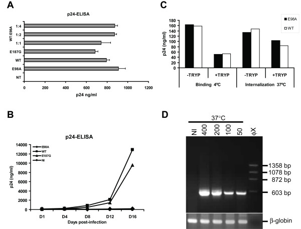
Virus release and internalization studies. p24-ELISA of transfected 293T cell (A) and infected H9 cell (B) culture supernatants. (A) 293T cells were transfected or co-transfected with mutant and wild-type pNL4-3 (2 μg) as indicated using the non-liposomal FuGENE transfection reagent (Roche) as recommended by the manufacturer. Culture supernatants were then assayed for p24 antigen contents 72 hrs post-transfection using an in-house p24 antigen ELISA [28]. Similar results were also obtained with transfected HeLa-tat cells. Virus stocks were then prepared from cleared and filtered culture supernatants (pre-cleared by centrifugation at 1,200 rpm for 7 min and filtered through a 0.45-μm-pore-size membrane) treated with DNase I (Roche) at 20 μg/ml final concentration at 37°C for 1 h. Aliquots in 300-μl fractions of the virus stocks were saved at -80°C until needed. (B) H9 cells (2 × 10⁵ cells) were infected with the X4 NL4-3 strain of mutant or wild type HIV-1 stocks using 200 ng of p24 antigen per well in 24-well plates. Three hours after infection, unbound viruses were removed by centrifugation, washed and resuspended in 1 ml complete RPMI medium per well. The infections were performed in triplicates and supernatants were collected at days 1, 4, 8, 12 and 16 post-infection and tested for p24 antigen contents by p24-ELISA. NI, non-infected control. (C) For virus binding and internalization assay, monolayered TZM-bl cells were seeded one day before infection and following day, medium was removed and cells were inoculated with equal amounts (400 ng of p24 antigen) of mutant or wild type NL4-3 virus stocks (treated with DNase I) with 20 μg/ml DEAE-dextran (in a total volume of 300 μl to 60,000 cells per well in 12-well plates). After adsorption period of 2 hrs, input viruses were removed and cells were treated with trypsin (+TRYP) or not (-TRYP) and the amount of cell associated p24 was measured using the p24-ELISA. (D) TZM-bl cells were also infected as described above with the amount virus indicated and after adsorption period of 2 hrs, input viruses were removed and cells were fed with 1 ml of complete DMEM with 5 μM indinavir and cultured for 24 hrs. Equal amounts of total RNA isolated from E98A infected TZM-bl cells were subjected to nested RT-PCR using specific primers that amplified a 593 bp fragment of the p17 viral RNA. The outer primer pair 5'-GCA GTG GCG CCC GAA CAG and 5'-TTCTGA TAA TGC TGA AAA CAT GGG TAT and inner primer pair 5'-CTC TCG ACG CAG GAC TC and 5'-ACC CAT GCA TTT AAA GTT CTA G was used. As an internal control, the human β-globin RNA was amplified using the primers described elsewhere [29].

