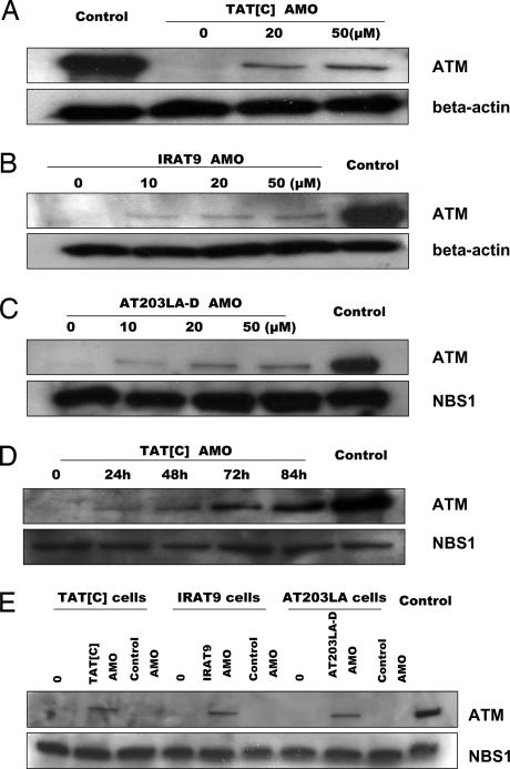
Fig. 3.
Immunoblots of nuclear lysates to demonstrate restoration of ATM protein levels to A-T cell lines after AMO treatment. Cells were treated for 24 h with different concentrations of cognate AMOs. Nuclear protein from normal cells was used as control. NBS1 or β-actin was used as a protein loading control. (A) TAT[C] cells were exposed to TAT[C]-AMO. (B) IRAT9 cells were exposed to IRAT9-AMO. (C) AT203LA cells were exposed to AT203LA-D AMO. (D) TAT[C] cells were exposed to 30 μM TAT[C]-AMO and harvested at different time points. (E) Specificity controls of AMOs. To evaluate the specificity of each AMO on protein expression, the AMOs were crisscrossed among the three cell lines. IRAT9-AMO was used as a control for TAT[C] and AT203LA cells; TAT[C]-AMO was used as a control for IRAT9 cells. For each cell line we treated cells with both the cognate AMO and a noncognate control AMO. No ATM protein was induced in cells exposed to optimized concentrations of noncognate AMOs (50 μM).

