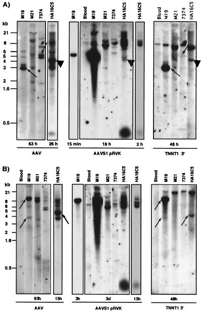
Figure 2.
wtAAV2 DNA integration can lead to the disruption of both AAVS1 and TNNT1. Southern blots of cellular DNA (ca. 10 μg) isolated from blood (negative control) and from cells latently infected with AAV (M19, M21, 7374, HA16) are shown. The genomic DNA was digested with either EcoRI (A) or HindIII (B). The blot was first hybridized to the 3′TNNT1 cDNA probe followed by hybridization to an AAV probe and subsequently to AAVS1pRVK. Between hybridizations, the blot was stripped (50% formamide, 2 h, 65°C) and the membrane was exposed to a PhosphorImager screen for 12 h to quantify remaining label. The exposure times are indicated for each blot. Cohybridization of AAV and TNNT1 is indicated by arrows (A, M19; B, M19, HA16) and of AAV, AAVS1, and TNNT1 by ▴ (A, HA16). Potential cohybridization of AAV and TNNT1 in 7374 cells was seen after EcoRI digestion (A); however, no TNNT1 disruption was apparent after HindIII digestion (B). Note, the variations in hybridization intensities with AAV as well as with AAVS1 probes are consistent with previous observations (see for example ref. 29)

