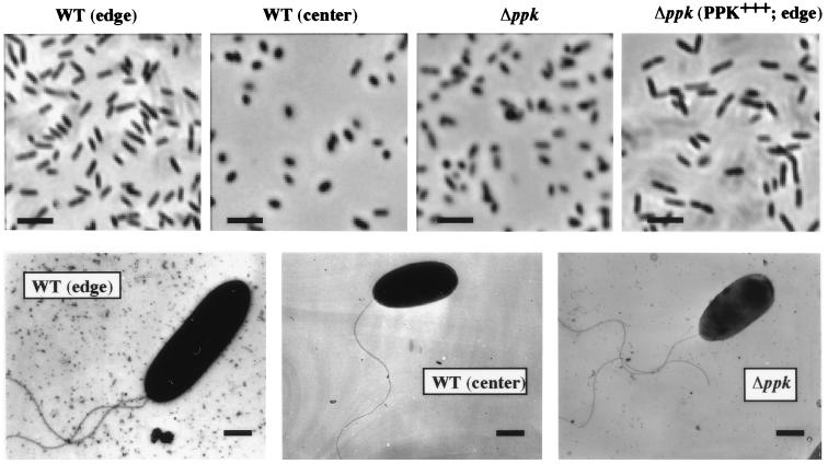
Figure 3.
Morphology of P. aeruginosa swarm cells by (Upper) phase-contrast and (Lower) electron microscopy. (Upper) Images of cells suspended in PBS from swarm colonies (X50 magnification). (Lower) Electron micrographs of bacteria taken directly from swarm plates (×10,400.) Strains: WT (edge), edge cells of PAO1 colony; WT (center), center cells of PAO1 colony; Δppk, cells from PAOM5 colony; and Δppk (PPK+++; edge), edge cells of PAOM5 colony containing pHEPAK11 plasmid. [Bars = 5 μm (Upper) and 0.5 μm (Lower).]

