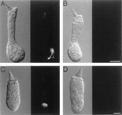
Figure 2.
Confocal images of immunolabeling with scFv-278 in isolated saccular hair cells. (A) In a hair cell shown by differential interference contrast microscopy (Left) to possess an immature hair bundle, immunoreactivity (Right) is concentrated in the perinuclear region of the cytoplasm and extends toward the apical cellular surface. (B) In a hair cell possessing an immature hair bundle, no immunolabeling occurs when scFv-278 is omitted. (The bar represents 5 μm in A and B.) (C) In a hair cell with a hair bundle of intermediate development, immunoreactivity occurs only in the perinuclear region. (D) A hair cell with a mature hair bundle displays no immunolabeling. (The bar represents 5 μm in C and D.)

