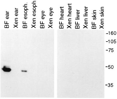
Figure 5.
Labeling of the cytokeratin on immunoblots of homogenates from bullfrog and Xenopus organs. Immunoblots of protein homogenates from bullfrog (denoted “BF”) and Xenopus (denoted “Xen”) organs reveal a single band recognized by the anti-cytokeratin scFv only in the bullfrog inner ear and esophagus. Molecular mass markers in kilodaltons are indicated at the right and apply to both blots.

