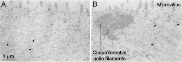
Figure 6.
Immunoelectron microscopic localization of the inner-ear cytokeratin to intermediate filaments in saccular cells. (A) Immunogold labeling of extramacular cells of the sacculus with the anti-cytokeratin scFv reveals numerous gold particles, three of which are indicated with arrowheads, on 10-nm filaments in the apical portion of the cell. Because of the extensive cellular permeabilization, many subcellular structures have been extracted; intermediate filaments and actin filaments persist. Note the lack of labeling of the microvilli located at the cellular apex. The scale bar pertains to both panels. (B) Within supporting cells, immunolabeling can be detected on cytoplasmic filaments located at the cellular apex; arrowheads demark the three gold particles in this section. Microvilli and the circumferential actin filaments associated with the junctional complex are unlabeled. In corroboration of the light microscopic observations, the immunoreactivity of supporting cells is weaker than that of extramacular cells.

