Figure 1.
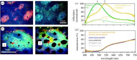
(a) The same iridophore ‘splotches’ viewed at near normal (red reflectance) and 45° incidence (blue–green reflectance) under white light illumination showing the spectral shift when increasing the angle of viewing on multilayer reflectors. (b) Iridophores can be covered within a fraction of a second by overlying, neurally controlled, pigmented chromatophore organs. Figure shows the same iridophore splotch with chromatophores retracted (1) and expanded (2). Arrows and numbers point to the area of spectral measurement shown in (c). (c) Normalized spectral reflectance measurements for both planes of polarization (parallel and perpendicular planes indicated by symbols) of iridophores only (green line peak at ca 480 nm) and yellow chromatophores covering the same iridophores (yellow line peak at ca 540 nm). Chromatophore pigments filter the wavelengths reflected from iridophores, yet linearly polarized light is not depolarized when passing upward through the chromatophore. (d) Transmission of light through yellow chromatophore, showing that chromatophores are neither birefringent nor optically active. Plot shows spectrum of unpolarized light transmitted through chromatophore (orange line), transmission spectrum of chromatophore placed between two polaroid filters in parallel (blue line) as well as when polaroids are crossed (90°; black line). Dark and light grey lines show chromatophore turned by 45° and 90°, respectively, while between crossed polaroids (cp). No transmission of light through cross-polarized, 45° (cp) and 90° (cp).

