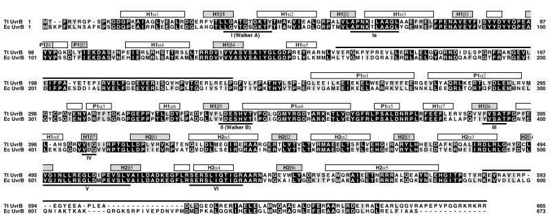
Figure 4.
Sequence alignment of Tt and Ec UvrB (30). Conserved residues are indicated by a black background. Similar residues (R/K, D/E, S/T, L/I/V/F/Y/M) have a gray background. Secondary structure elements are indicated above the sequences (α-helices are represented by white boxes, β-strands by gray boxes) and are numbered sequentially (first two letters denote the corresponding domain). The regions that are disordered in the crystal structure are marked by a black line above the sequences. Helicase consensus sequences (39) are labeled with Roman numbers.

