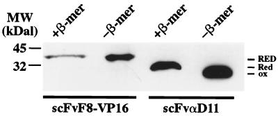
Figure 5.
Redox state of scFv fragments. Western blot analysis of scFvF8-VP16 (expressed in the cytoplasm of L40 yeast cells) and of a secreted scFv fragment (scFvαD11, expressed as a secreted protein in the endoplasmic reticulum of insect cells). Samples were prepared in SDS/PAGE loading buffer containing (+ β-mer) or not (− β-mer) β-mercaptoethanol. After blotting, the scFv fragments were detected with the anti-myc antibody 9E10. The bars at the right of the lanes indicate the molecular mass gel shift between the oxidized (ox) and reduced (red) forms of the scFvαD11. ScFvF8-VP16 fusion protein does not show a shift in electrophoretic mobility, between reducing and nonreducing conditions, indicating that it does not form disulfide bonds in the yeast cytoplasm (RED).

