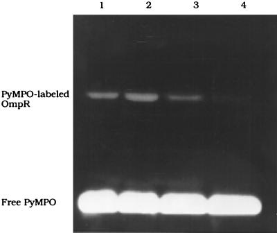
Figure 4.
Fluorescent labeling of OmpR with PyMPO. OmpR (9.5 μM) is reacted with a 50-fold excess of PyMPO for 30 min at RT in 40 mM Imidazole/1 mM EDTA, pH 7.6 (lane 1). Lane 2 shows a stimulation of PyMPO labeling in the presence of C1. OmpR (9.5 μM) is incubated with acetyl phosphate for 3 h at RT in the absence (lane 3) or presence (lane 4) of C1 DNA and then labeled with PyMPO as before. The products of the labeling reaction are separated on SDS/12% PAGE. The visible fluorescent band is the PyMPO-labeled OmpR protein.

