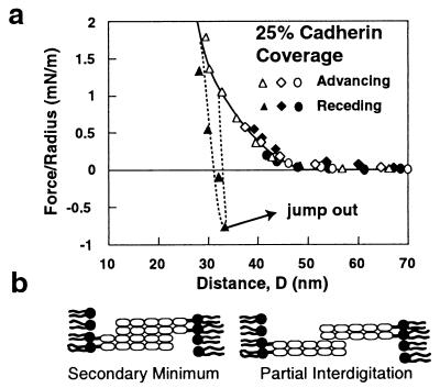
Figure 4.
(a) “Partial interdigitation” measurements obtained with cadherin monolayers at 25% surface coverage. Successive measurements correspond with successively decreasing intermembrane separations D. The circles indicate the forces measured during the approach ( ) and retraction ( ) of the protein films for the closest intermembrane distance of 40 nm. The diamonds indicate a successive measurement in which the distance of closest membrane approach was 37 nm, and the open and filled triangles indicate advancing and receding measurements, respectively, to and from 29 nm. The solid arrow shows the jump-out from the adhesive minimum at 32 nm. (b) The proposed protein alignments at partial protein interdigitation.

