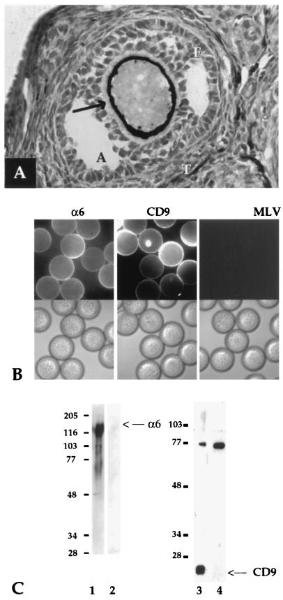
Figure 1.
Expression of α6 and CD9 on the surface of oocytes and mature, fertilization-competent eggs (A). Ovarian sections from mature mice were processed and stained for CD9. A secondary follicle is shown. Arrow indicates staining with the mAb KMC 8.8 on the oocyte plasma membrane and fine fibrillar staining extending into the zona pellucida. T, thecal layer; F, follicular cells; A, antrum. Staining was also seen on oocytes in Graafian follicles (data not shown). (B) Mature zona-free mouse eggs were incubated with GoH3 (Left), JF9 (Center), or a control rat mAb, anti-MLV (Right), and processed for immunofluorescence; fluorescence (Upper) and phase-contrast (Lower). (C) Zona-free biotinylated mouse eggs were lysed and processed for immunoprecipitation with anti-α6 mAb, GoH3 (lane 1), or anti-CD9 mAb, JF9 (lane 3). Samples were first immunoprecipitated with an anti-virus mAb, anti-MLV (lanes 2 and 4). Samples were analyzed by nonreducing 10% SDS/PAGE. We found no evidence (by Western analysis or immunofluorescence) for CD9 expression on mature sperm (data not shown).

