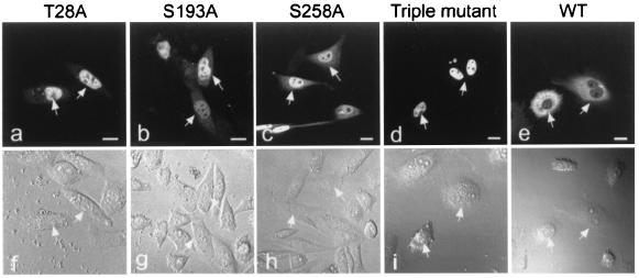
Figure 3.
Expression of AFX-GFP mutants in CHO-K1 cells. CHO-K1 cells were transfected with either the wild-type AFX-GFP (WT) or each mutant, as indicated at the top, and the fluorescence of the proteins (a–e) and morphology of the cells observed under a Nomarski interfering microscope (f–j) are shown. The positions of the nuclei of the cells expressing AFX-GFP are indicated by arrows. (Bars, 10 μm.)

