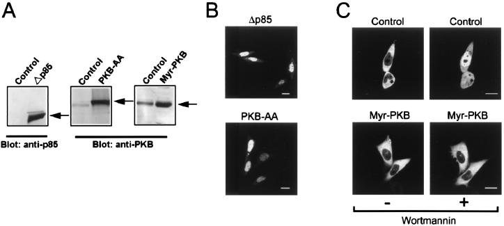
Figure 4.
Localization of AFX-GFP in CHO-K1 cells infected with the dominant-negative PI 3-kinase, the dominant-negative PKB, and the active form of PKB. (A) Immunoblot analysis of PI 3-kinase and PKB. The lysates prepared from the cells infected with the adenovirus vectors encoding the dominant-negative PI 3-kinase (Δp85), the dominant-negative PKB (PKB-AA), the active form of PKB (Myr-PKB), or β-galactosidase (Control) were subjected to immunoblot analysis using the anti-p85 subunit of PI 3-kinase Ab or the anti-PKB Ab. The positions of the immunoreactive bands are indicated by arrows. (B and C) Localization of AFX-GFP in infected cells. CHO-K1 cells expressing AFX-GFP were infected with each adenovirus vector as indicated at the top, and the fluorescence of AFX-GFP was observed. (C) The localization of AFX-GFP was observed before and after treatment with wortmannin at 1 μM for 30 min. (Bars, 10 μm.)

