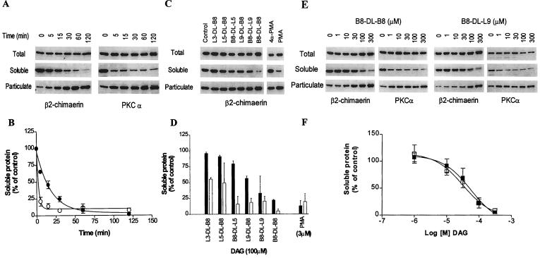
Figure 2.
Translocation of β2-chimaerin by DAG lactones. COS-1 cells were transfected with pCR3ɛ-β2-chimaerin and 48 h later were treated with cyclic DAGs. Soluble and particulate fractions were separated by ultracentrifugation and subjected to Western blot analysis using the anti-ɛ-tag antibody (for ɛ-tagged β2-chimaerin) or anti-PKCα antibody, as indicated. (A, C, and E) Representative Western blots. The molecular mass of β2-chimaerin is 50 kDa and that of PKCα is 80 kDa. (B, D, and F) The densitometric analysis of the immunoreactivity in the soluble fraction for A, C, and E, respectively. Results are expressed as percentage of the values observed in control cells and represent the mean ± SE of three independent experiments. (A and B) Time course of translocation after treatment of COS-1-transfected cells with B8-DL-B8 (100 μM); β2-chimaerin (●); PKCα (○). (C and D) Translocation of β2-chimaerin by different cyclic DAGs (100 μM) after 1 h treatment; β2-chimaerin (solid bars); PKCα (open bars). (E and F) Concentration-dependence of β2-chimaerin translocation after 1 h incubation with B8-DL-B8 (■) or B8-DL-L9 (□).

