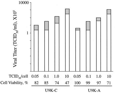
Figure 2.
Cell viability and viral release from EMCV-infected cells. Cells (0.5 × 10⁶) were infected with EMCV with doses from 0.05 to 10 TCID50/cell for 12 hr. After harvesting, viral titers in the cell pellets (freeze-thawed three times) and supernatants were measured by TCID50 bioassays on L-929 cells; cell viability was determined by trypan blue viable cell counts. Viral titers are plotted on a log10 scale. For each stacked bar, the upper and lower portions correspond to EMCV titers in the pellet and supernatant, respectively.

