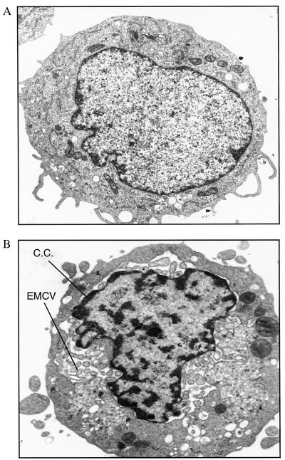
Figure 3.
Electron microscopy of U9K-A cells persistently infected with EMCV. Noninfected U9K-A controls and U9K-AV2 cells persistently infected with EMCV were examined with transmission electron microscopy for the presence of viral particles. (A) Control U9K-A cells. (B) U9K-AV2 cells. Magnification: ×16,000. C.C., chromatin condensation.

