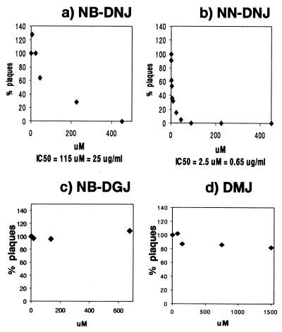
Figure 2.
Secretion of infectious BVDV in the presence of N-butyl-DNJ (a), N-nonyl-DNJ (b), N-butyl-DGJ (c), and DMJ (d). (a and b) MDBK cells were grown to confluency in six-well plates and infected with 500 pfu of cp BVDV (NADL strain) per well for 1 hr at 37°. The inoculum then was replaced with medium containing the indicated concentrations of either NB-DNJ, NN-DNJ, NB-DGJ, or DMJ (plaque assay). After 3 days the supernatants were removed and used to infect fresh MDBK monolayers in six-well plates. The presence of inhibitor during the 1-hr infection does not have an effect (data not shown). After 1 hr the inoculum was removed and the cells were washed thoroughly and incubated with inhibitor-free medium. After 3 days the plaques were counted (yield assay) and the results were expressed as a percentage of the number of plaques resulting from infection with the inhibitor-free plaque assay supernatant (=100%) (x axis). The y axis indicates the inhibitor concentrations used in the plaque assay. The IC50 is indicated at the bottom. (c and d) Plaque assay results are shown. NB-DGJ was used at concentrations of up to 680 μM, an inhibitor concentration sufficient to inhibit completely the ceramide-specific glucosyltransferase (data not shown). DMJ was used at concentrations of up to 1.5 mM, an inhibitor concentration sufficient to protect treated cells from killing by a complex sugar-binding lectin (ECA) (data not shown). All experiments were done at least in duplicate, and each data point represents the average of two wells counted. Experiment 2 (b) was done in triplicate (n = 3 ± 0.13), and each data point represents the average of two wells counted.

