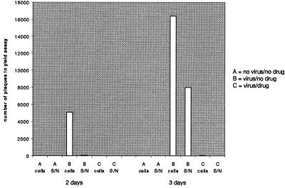
Figure 4.
Infectivity of viral material inside and outside of NB-DNJ-treated cells 2 and 3 days after infection. Noninfected (A) and BVDV-infected (B and C) MDBK cells were grown in the absence (A and B) or presence (C) of 1 mg/ml NB-DNJ for either 2 or 3 days. The supernatants were saved and the cells were washed and lysed by freeze-thawing. Yield assays were performed to determine the number of pfus in the supernatants (S/N) and cell lysates (cells).

