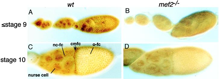
Figure 2.
D-MEF2 expression in developing egg chambers. Ovaries were dissected from either Oregon-R (wt; A and C) or D-mef265/D-mef2424 (mef2−/−; B and D) and stained with an anti-D-MEF2 polyclonal antibody. Anterior is to the left. (A and B) Stage 9 and early eggs. (C and D) Stage 10 eggs. In wild type, D-MEF2 is detected in the nuclei of both nurse and follicle cells up to stage 13 (later stages not shown). Nuclear staining of D-MEF2 is reduced in the mutant background. nc-fc, nurse cell-associated follicle cells; cmfc, centripetally migrating follicle cells; o-fc, oocyte-associated follicle cells.

