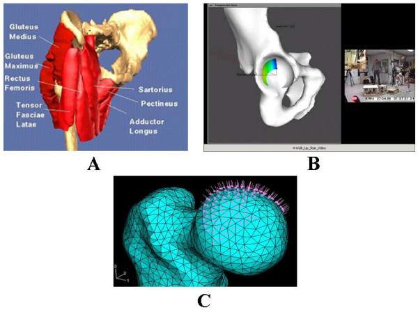
Figure 4.
(A). The surface model of the pelvis and the proximal femur with the key muscles across the joint used for the dynamic force analysis of the hip. (B). The model used to study acetabulum contact area and stress distribution during activities of daily living involving the hip. The hip joint reaction force (arrow) and contact stress distribution at three positions during the gait cycle for the left (highlighted) leg calculated using the discrete element analysis (DEA) technique. The blue areas indicate the regions of the lowest stress while the yellow and green regions indicate the locations of higher stresses. (C). The proximal femur model used to investigate subchondral bone collapse due to osteonecrosis (OS) and femoral head reconstruction.

