
Figure 4.
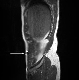
In-vivo localisation of the gold acupuncture needle inserted into the Dai mai point as seen in a saggital slice reconstruction. The white arrow simulates the direction of needle insertion. Image reconstruction was done with the J-Vision software of the TIANI workstation. MRI acquisition data: TR = 721, TE = 19, flip = 150, TH = 3, TF = 3, FOV = 370, MA = 320.