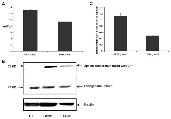
Figure 4.

Difference in mRNA and protein expression levels between c.602C and c.602T variants in OPTC. (A): Quantiative RT-PCR showing expression of c.602C and c.602T in mRNA level. The difference in mRNA expression is represented by ΔΔCT value. (B): Western blot of two variants of Opticin core protein fused with GFP as well as endogenous Opticin in RPE cells. UT represents the untransfected RPE cells. (C): Bar diagram showing the mean ± SD of densitometric scanning of western blots done thrice, that indicates much less expression of OPTC-c.602T variant (43%) than the normal one (OPTC-c.602C). Expression of transfected protein (Opticin-GFP) was normalized with endogenous Opticin.
