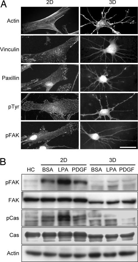
Fig. 3.
Fibroblasts interacting with relaxed collagen matrices lack stress fibers, focal adhesion structures, and focal adhesion signaling. (A) Fibroblasts were incubated 4 h in medium containing 50 ng/ml PDGF on collagen-coated coverslips (2D; Left) or on top of relaxed collagen matrices (3D; Right) after which samples were fixed and stained for actin or focal adhesion proteins. Cells on coverslips but not matrices formed stress fibers and focal adhesions containing vinculin, paxillin, pTyr, and pFAK. (Scale bar: 50 μm.) (B) Fibroblasts were incubated 4 h in medium with or without 50 ng/ml PDGF or 10 μM LPA as indicated on collagen-coated coverslips (2D; Left) or inside relaxed collagen matrices (3D; Right) after which samples were extracted and subjected to immunoblotting for pFAK, FAK, p130Cas (Cas), phospo-Cas (pCas), and actin. Compared with freshly harvested cells (HC), fibroblasts incubated on coverslips but not within relaxed collagen matrices showed FAK and Cas activation (phosphorylation).

