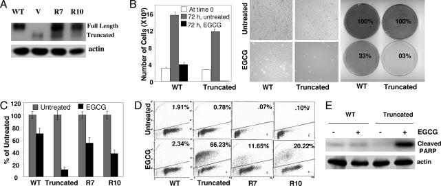
Fig. 1.
SHP-2 negatively regulates EGCG-induced apoptosis. (A) Total cell lysates from cells expressing WT or truncated SHP-2 and two rescued clones (R7 and R10) were immunoblotted with anti-SHP-2. (B) Cells were treated with 120 μM EGCG for 72 h. Live cell numbers were counted by the trypan blue dye exclusion method, and plates were photographed and stained with methylene blue. (C) Cells were treated with EGCG for 72 h. Five hundred live cells were plated for colony formation. Results are plotted as percentages of untreated controls. (D) Cells were treated with EGCG for 72 h, fixed in ethanol, and stained with Apo-BrdU to determine the apoptotic population. (E) Cells were treated with EGCG for 24 h, and total cell lysates were immunoblotted with an antibody that specifically detects cleaved PARP (89 kDa) only.

