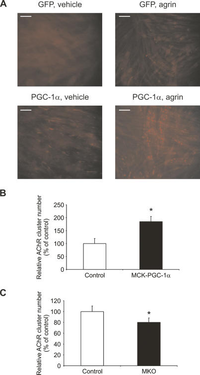
Figure 2.
AChR clustering induced by PGC-1α. (A) C2C12 myotubes were infected with adenoviral GFP and PGC-1α, respectively, for 48 h. Sixteen hours before harvesting, the myotubes were treated with vehicle (PBS) or 10 ng/mL recombinant agrin. Subsequently, cells were fixed and incubated with rhodamine-labeled α-BTX, and AChR clusters were visualized by fluorescence microscopy. (B,C) The average number of NMJ counted in 20 fields per genotype was normalized to the number of nuclei in these fields, and relative acetylcholine cluster numbers were expressed as percentage of control mice. (*) p < 0.05 between muscle fibers from MCK-PGC-1α and controls as well as fibers from MKO and controls, respectively.

