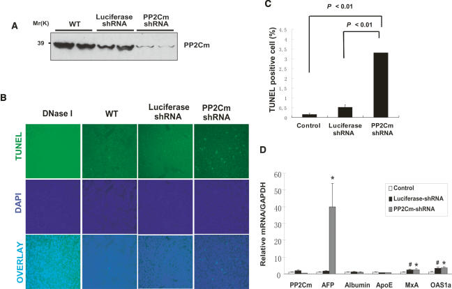
Figure 5.
Loss of PP2Cm in vivo induces hepatocyte apoptosis and liver injury. (A) Western blot analysis of PP2Cm expression in total liver lysates from wild-type mice or mice treated with adenovirus expressing either luciferase control shRNA or PP2Cm-specific shRNA3 for 7 d. (B, top panel) Mouse livers treated as described above were fixed and assessed for apoptisis by TUNEL staining. (Middle panel) The nuclei were counterstained with DAPI. (Bottom panel) Corresponding images were overlaid to distinguish false positive signals. DNase I-treated liver sections served as a positive control. (C) The percentage of apoptotic cells was tallied and presented as mean ± SD (n = 4). (D) Hepatic gene profiles of mouse livers treated as above were examined with quantitative RT–PCR by normalizing against GAPDH. (*) p < 0.05, PP2Cm shRNA versus control; (#) p < 0.05, luciferase shRNA versus control, Student’s t-test.

