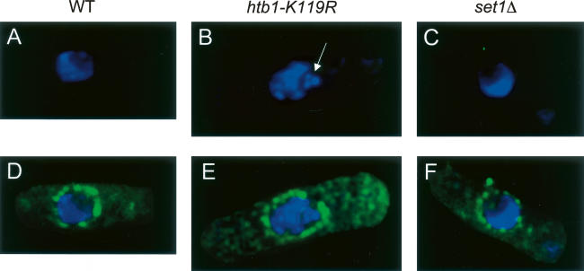
Figure 4.
Abnormal nuclear morphology in the htb1-K119R mutant. Wild-type, htb1-K119R, and set1Δ cells were stained with a nuclear pore antibody and DAPI. Shown are deconvolved images taken using a DeltaVision microscope equipped with a CCD camera. (A–C) DAPI stain alone. (D–F) DAPI stain merged with nuclear pore stain. The white arrow in B denotes a fragment of nuclear, DAPI-stained material in the htb1-K119R mutant cell.

