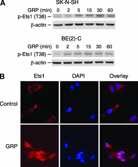
Figure 3.
Ets1 phosphorylation and nuclear localization after GRP treatment. (A) SK-N-SH cells were serum-starved overnight and then treated with GRP (10-7 M). Induction of Ets1 phosphorylation was detected after GRP treatment in both SK-N-SH and BE(2)-C cells by Western blot analysis. (B) For immunofluorescent staining, SK-N-SH cells were seeded on coverslips and serum-starved overnight. Ets1 protein was localized predominantly in the nuclei at 1 hour (bottom panels) after GRP treatment (10-7 M) when compared to controls (top panels). DAPI specifically stained for the DNA of nuclei.

