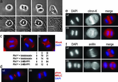
Fig. 4.
Acute inhibition of Plk1 during anaphase disrupts RhoA localization and blocks the onset of cytokinesis. (a and b) Metaphase Plk1wt (a) and Plk1as (b) cells were treated with 10 μM 3-MB-PP1 (time 0) and followed during progression into anaphase. Although chromatid separation occurred normally, Plk1as cells failed to divide and became binucleated (SI Movies 1 and 2). (c) Plk1 activity is required to localize RhoA at the equatorial cortex. Plk1wt and Plk1as cells were synchronized with monastrol, released for 30 min, and then treated for 20 min with either blebbistatin or 3-MB-PP1. RhoA accumulation at the equatorial cortex was visualized after trichloroacetic acid fixation (21, 27). Anaphase cells (n = 100 per sample) were classified with respect to equatorial RhoA staining and cleavage furrow formation. (d–f) Plk1wt and Plk1as cells were treated with 3-MB-PP1 and stained with antibodies against phosphorylated myosin regulatory light chain (MRLC) (d), citron kinase (citron-K) (e), and anillin (f).

