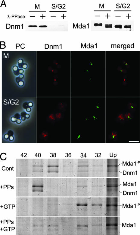
Fig. 5.
Phosphorylation of Mda1. (A) The purified proteins from M-arrest or S/G2-arrest were treated with (+) or without (−) λ-protein phosphatase (λ-PPase), subjected to low-bis-acrylamide gel electrophoresis and immunoblotted with anti-Dnm1 or anti-Mda1. (B) Cells in M-arrest or S/G2-arrest were immunostained with anti-Dnm1 and anti-Mda1 antibodies and detected with Alexa Fluor 555- or Alexa Fluro 488-conjugated secondary antibody, respectively. (Scale bar: 2 μm.) (C) Narrow-ranged differential density centrifugation of Mda1-containing macromolecules in various states. Partly purified fraction from M-arrest cells was incubated in the presence of phosphatase and/or GTP, then fractionated by differential density centrifugation, and analyzed by SDS/PAGE. Each lane contains pellets through a certain concentration of iodixanol (%) as indicated above. Cont, incubated in control buffer; +PPs, incubated in the presence of λ-PPase; +GTP, incubated in the presence of GTP; Mda1-p, band for natively phosphorylated Mda1; Mda1, band for Mda1 dephosphorylated in vitro.

