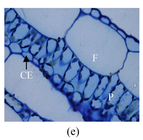
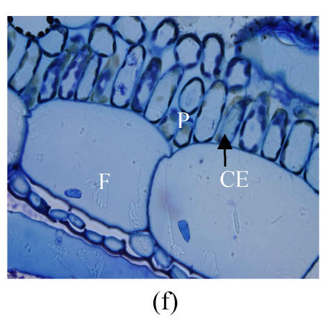
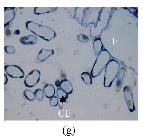
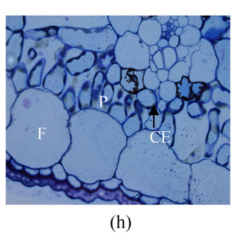
Fig. 1.
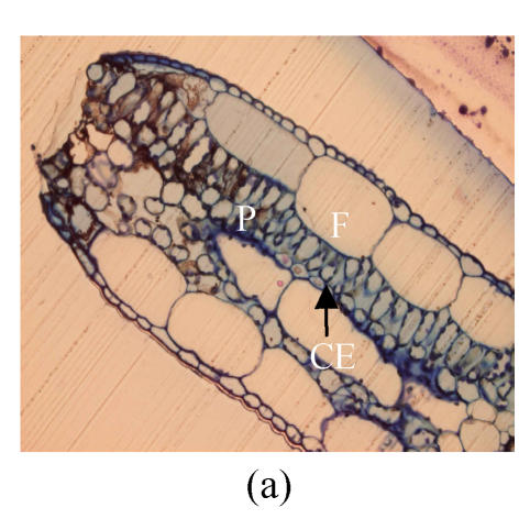
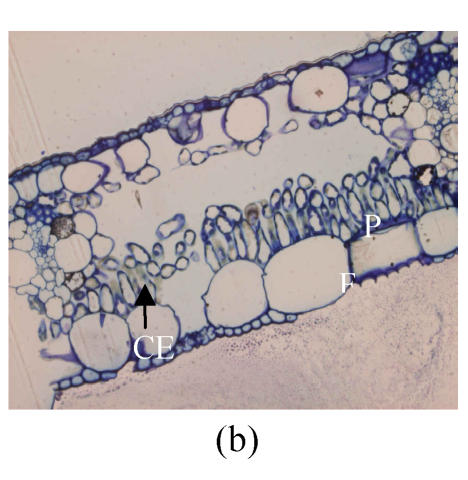
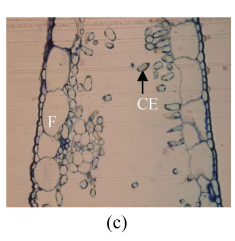
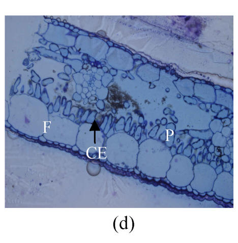
Light microscopy observation for effects of SA on cell structure in chilling-stressed banana seedling leaves. (a) Mesophyll cells of banana seedlings growing at 30/22 °C (10×40); (b) Mesophyll cells of banana seedlings treated by 0.5 mmol/L SA at 30/22 °C for 1 d (10×40); (c) Mesophyll cells of banana seedlings suffered from 7 °C stress for 3 d (10×40); (d) Mesophyll cells of SA-pretreated banana seedlings suffered from 7 °C stress for 3 d (10×40); (e) Mesophyll cells of banana seedlings growing at 30/22 °C (10×100); (f) Mesophyll cells of banana seedlings treated by 0.5 mmol/L SA at 30/22 °C for 1 d (10×100); (g) Mesophyll cells of banana seedlings suffered from 7 °C stress for 3 d (10×100); (h) Mesophyll cells of SA-pretreated banana seedlings suffered from 7 °C stress for 3 d (10×100)
CE: Cell; F: Fibro; P: Palisade parenchymas

