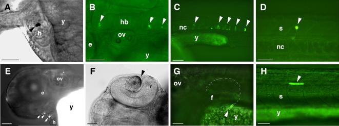
Figure 4.
Tissue Type Specific Expression of GFP Reporter Gene in Zebrafish Embryos. Examples of GFP expression induced by CNEs 1, 9, 10, and 11 are shown in fixed tissues after wholemount anti-GFP immunostaining (bright field views A and F) or in live embryos by combined bright field and GFP fluorescence microscopy analyses (B, C, D, E, G and H). Arrowheads indicate GFP expressing cells. Embryos C and D are ∼26–33 hpf, while embryos A, B, E, F, G, and H are 48–54 hpf. Lateral views, anterior to the left and dorsal to the top except for F where the dorsal view is shown. GFP positive cells were found in the following: (A) CNE1, heart chamber (B) CNE1, hindbrain neurons (C) CNE9, notochord (D) CNE9, spinal cord neuron (E) CNE10, lower jaw primordia and pericardial regions (F) CNE10, lens epithelial cell layer (G) CNE11, pectoral fin (H) CNE11, muscle. (e) Eye; (f) fin; (h) heart; (hb) hindbrain; (I) lens; (nc) notochord; (ov) otic vesicle; (r) retina; (s) spinal cord; (y) yolk.

