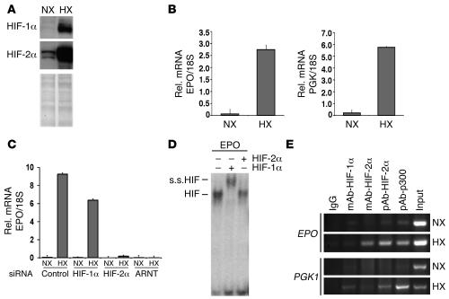
Figure 7. HIF-2α preferentially binds to the endogenous EPO 3′ HRE in hepatocytes.
(A) Western blot analysis for HIF-1 and HIF-2 in normoxic (21% O2; NX) and hypoxic (1% O2; HX) Hep3B cells. (B) EPO and PGK1 mRNA levels in normoxic and hypoxic Hep3B cells as determined by real-time PCR. (C) The hypoxic induction of EPO expression in Hep3B cells is HIF-2 dependent. Real-time PCR analysis of EPO expression in normoxic and hypoxic Hep3B cells treated with control, HIF-1α, HIF-2α, or control siRNA oligonucleotides. Error bars represent SD. (D) HIF-1α preferentially binds to the EPO HRE in vitro as determined by EMSA. s.s.HIF, HIF supershift. + and – indicate the presence and absence, respectively, of the antibody used in a supershift reaction. (E) ChIP analysis of the EPO and PGK1 HREs in normoxic and hypoxic Hep3B cells using antibodies directed against HIF-1α, HIF-2α, and CBP/p300. Coprecipitated DNA fragments were detected by PCR using primers spanning the EPO and PGK1 HREs. mAb-HIF-1α, mAb against HIF-1α; pAb-HIF-2α, polyclonal antibody against human HIF-2α; pAb-p300, polyclonal antibody against human p300.

