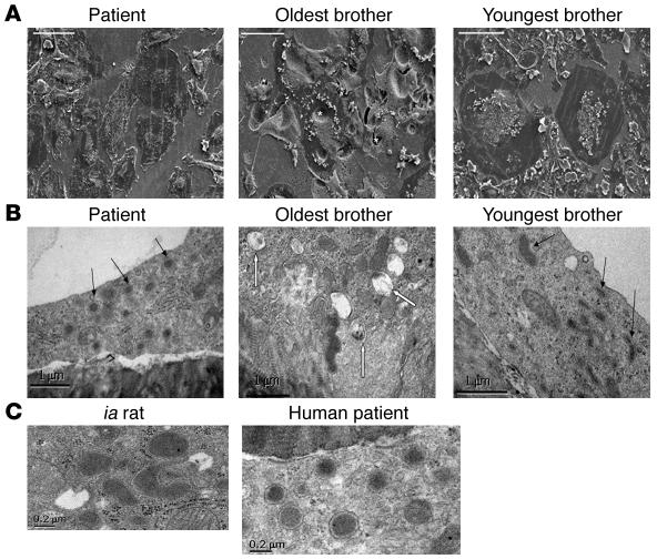
Figure 5. Electron microscopy of osteoclasts generated from the family with autosomal-recessive osteopetrosis.
(A and B) Osteoclasts were formed from all 3 siblings, then fixed and analyzed by scanning EM or transmission EM. (A) Scanning EM images show that the oldest brother produced normal, actively resorbing osteoclasts excavating deep resorption pits (denoted by asterisks), whereas both the patient and the youngest brother formed large, very flat osteoclasts with little evidence of resorption. (B) Transmission EM demonstrated that both the patient and the youngest brother produced osteoclasts containing large numbers of electron-dense granules (black arrows), whereas the resorbing osteoclasts formed from the oldest brother contained many multivesicular bodies (white arrows). (C) The electron-dense vesicles were identical in ultrastructure to those seen in the ia rat, which are known to contain TRAP. Scale bars: 100 μm (A), 1 μm (B), 0.2 μm (C).

