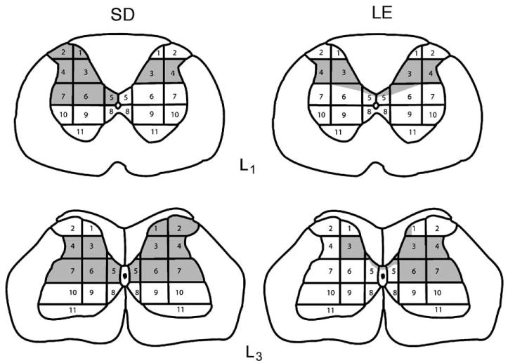
Fig. 1.

Schematic diagrams show the area of tissue damage for representative sections from SD and LE spinal-injured animals. The 11 regions of spinal gray matter are the same as those used in a previous study to examine the morphological correlate of spontaneous and evoked pain behaviors following QUIS injections (Yezierski et al., 1998).
