
Fig. 6.
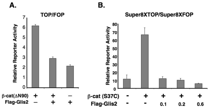
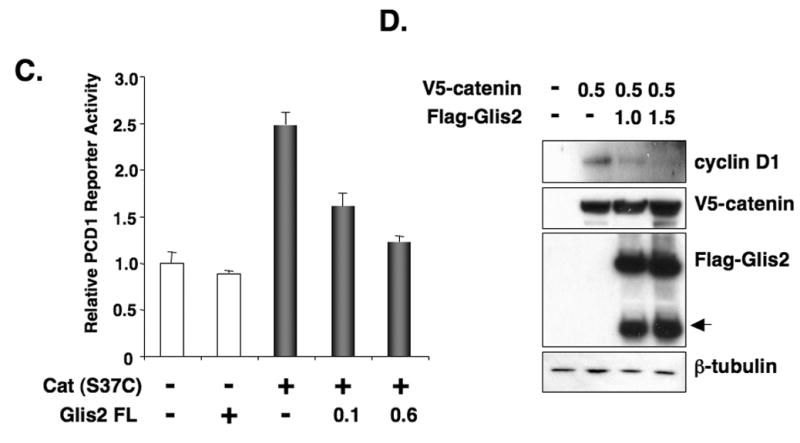
Glis2 represses TCF-mediated transcriptional activity and the induction of cyclin D1 by β-catenin. (A) HCT116 cells were cotransfected with TOPFlash and FOPFlash reporters, pCMVbeta, β-catenin(ΔN90), and p3xFlag-CMV-Glis2 as indicated. Forty-eight h after transfection, cells were assayed for reporter activities as described in Materials and Methods. (B) HCT116 cells were cotransfected with Super8XTOPFlash and Super8XFOPFlash reporters, pRL-SV40, pCMV-V5-β-catenin (β-cat(S37C)) and different concentrations of p3xFlag-CMV-Glis2 expression vector as indicated and then processed as described under A. (C) HCT116 cells were cotransfected with the cyclin D1 reporter PCD1, pRL-SV40, the constitutively-active pCMV-V5-β-catenin (S37C) expression vector, and p3xFlag-CMV-Glis2 as indicated and then processed as described under A. (D) HCT116 cells were transfected with pCMV-V5-β-catenin and p3xFlag-CMV-Glis2 (1.0 and 1.5 μg) as indicated. Thirty-six h later, cellular proteins were analyzed by Western blot analysis using antibodies against V5, Flag, β-tubulin, and cyclin D1. The lower molecular weight band (arrow head) in the Flag-Glis2 Western blot represents a proteolytically processed form of Glis2.