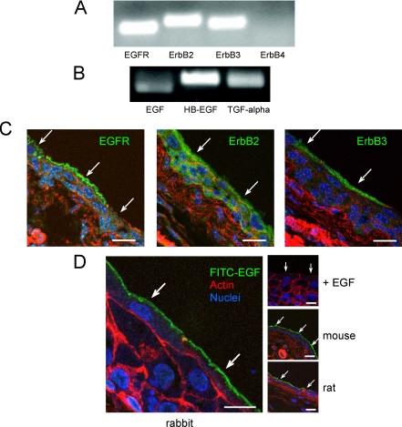
Figure 3.
Expression and distribution of ErbB family receptors in the uroepithelium. (A and B) Total RNA was prepared from rabbit uroepithelium and RT-PCR used to assess expression of ErbB1–4 (A) or EGFR ligands EGF, HB-EGF, and TGFα (B). (C) Localization of ErbB family receptors in cryosections of mouse uroepithelium, labeled with antibodies to visualize the receptors (green), rhodamine-phalloidin to label the actin cytoskeleton (red), and Topro-3 to label nuclei (blue). Bar, 20 μm. (D) Binding of FITC-EGF to rabbit uroepithelium (far left), in which tissue was bathed in 40 ng/ml FITC-EGF at 4°C for 1 h, washed, fixed, and sectioned. In the control study (right, top), FITC-EGF binding to rabbit tissue was competed with excess unlabeled EGF (400 ng/ml). FITC-EGF binding to the apical surface of mouse and rat umbrella cells is shown in the right middle and right bottom panel, respectively. Bar, 15 μm for rabbit tissues and 20 μm for mouse and rat tissues. Individual umbrella cells are indicated by arrows.

