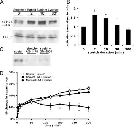
Figure 5.
Stretch activates the EGFR. (A) Tissue was stretched for the indicated time, lysates of rabbit uroepithelium were prepared and resolved by SDS-PAGE, and Western blots were probed with antibodies specific for phosphorylated Y1173-EGFR or total EGFR. (B) Quantification of Y1173 phosphorylation in response to stretch, relative to unstretched (t = 0′) tissue samples. The mean changes in capacitance ± SEM are shown (n = 4). *, Statistically significant difference (p < 0.05) relative to unstretched tissue. (C) Tissue was stretched for 2 min in the absence of additional treatment, or pretreated for 30 min with 25 nM AG-1478 or 10 μM GM-6001 before stretch, followed by lysate preparation. (D) Rabbit uroepithelium was placed on tissue rings and the mucosal or serosal surfaces were pretreated with 1 μg/ml LA1 EGFR function-blocking antibody for 1 h before mounting, equilibrating, and stretching the tissue in Ussing stretch chambers. The mean changes in capacitance ± SEM (n ≥ 3) are shown. *, Statistically significant difference (p < 0.05) relative to control stretch samples.

