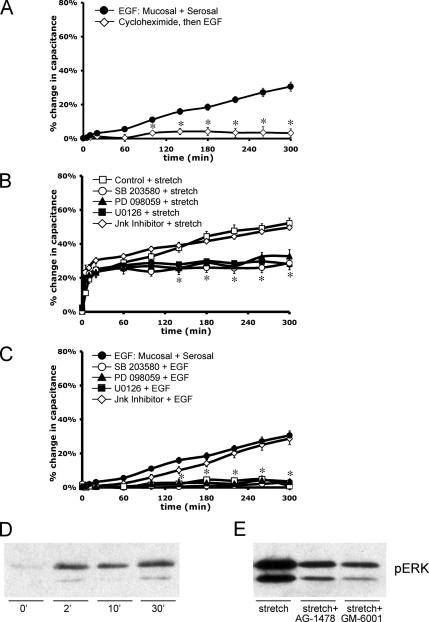
Figure 7.
Protein synthesis and MAPK signaling pathways are required for EGF- and stretch-induced late-phase capacitance increases. Rabbit uroepithelium was isolated and mounted in Ussing stretch chambers. (A) Tissue was incubated with 100 ng/ml EGF (±60-min pretreatment with 100 μg/ml cycloheximide). *, Statistically significant difference (p < 0.05) relative to EGF-treated samples. (B) Before stretch, tissue was pretreated with p38 inhibitor SB-203580 (10 μM for 1 h), MEK1 inhibitor PD-098059 (10 μM for 30 min), MEK1/2 inhibitor U0126 (10 μM for 30 min), or JNK inhibitor II (500 nM for 30 min) as indicated. *, Statistically significant differences (p < 0.05) relative to control samples were observed for tissue treated with SB-203580, PD-098059, or U0126 at t ≥140 min. (C) Before treatment with 100 ng/ml EGF, tissue was incubated with SB-203580, PD-098059, U0126, or JNK inhibitor II as described above. *, Statistically significant differences (p < 0.05) relative to control samples were observed for tissue treated with SB-203580, PD-098059, or U0126 at t ≥100 min. In each panel, the mean changes in capacitance ± SEM (n ≥ 3) are shown. (D) Tissue was stretched for the indicated time before lysate preparation. Western blots were probed with antibodies specific for pERK1/2. (E) Tissue was left untreated (stretch) or pretreated for 30 min with 25 nM AG-1478 (AG) or 10 μM GM-6001 (GM) as indicated. Tissue was then stretched for 2 min before lysate preparation and blotting with phospho-specific antibodies to ERK1/2.

