Figure 2.
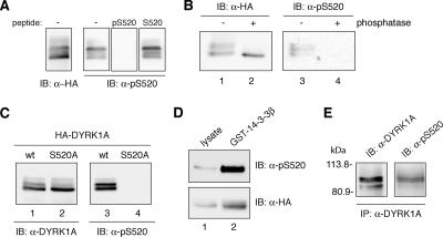
DYRK1A is phosphorylated on Ser-520 in vivo. (A) Whole-cell extracts from U2-OS cells expressing HA-DYRK1Awt were resolved by 8% SDS-PAGE and analyzed by immunoblot with anti-HA antibody (left) and with anti-pS520 antibody (right). Where indicated, the phosphorylated immunizing peptide (pS520: SNSGRARpSDPTHQHR) or an equivalent unphosphorylated peptide (S520: SNSGRARSDPTHQHR) was added at 25 μg/ml during the incubation period with 1 μg/ml anti-pS520 antibody. (B) Lysates from U2-OS cells transiently transfected with pHA-DYRK1A were incubated for 30 min at 30°C in phosphatase buffer alone (lanes 1 and 3) or supplemented with alkaline phosphatase (lanes 2 and 4), as indicated. Samples were analyzed by SDS-PAGE and immunoblot with anti-HA antibody (lanes 1 and 2) and with the anti-DYRK1A pS520 antibody (lanes 3 and 4). (C) U2-OS cells were transiently transfected with pHA-DYRK1Awt (lanes 1 and 3) and pHA-DYRK1A/S520A (lanes 2 and 4). Whole-cell extracts were resolved by 8% SDS-PAGE and immunoblotted with an anti-HA antibody (lanes 1 and 2) and with the anti-DYRK1A pS520 antibody (lanes 3 and 4). (D) Soluble cell extracts from U2-OS cells expressing HA-DYRK1Awt were incubated with GST-14-3-3β. Lysates, representing 10% of the input, (lane 1) and pulled-down proteins (lane 2) were separated by 8% SDS-PAGE and analyzed by immunoblot with anti-HA antibody (bottom) and with the phospho-specific anti-DYRK1A pS520 antibody (top) to detect the presence of Ser-520–phosphorylated DYRK1A bound to 14-3-3β. (E) The endogenous DYRK1A protein was immunoprecipitated from PC12 cells with anti-DYRK1A antibody. The immunoprecipitate was analyzed by immunoblot (8% SDS-PAGE) with both the anti-DYRK1A and the anti-DYRK1A pS520 antibody. The position of marker proteins (in kilodaltons) is indicated.

