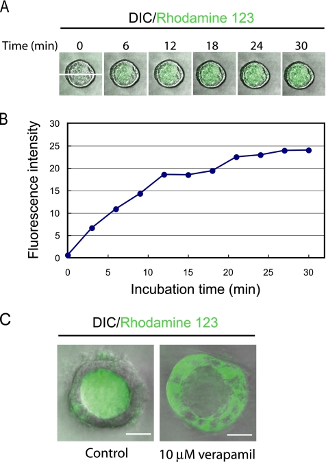
Figure 4.
HPPL cysts transport rhodamine 123 from the basal side to the apical. (A) HPPL cysts accumulated rhodamine 123 in the central lumen. HPPL cysts formed in 40% Matrigel were incubated with rhodamine 123, an mdr substrate, for 5 min. After washing the culture, time-lapse images were taken by a confocal microscope. (B) The time course of transport of rhodamine 123. HPPL transported rhodamine 123 efficiently during the first 15 min into the central lumen, and then the fluorescence intensity inside the lumen almost reached a plateau after 20 min. The fluorescence intensity values along the x-y axis shown in the picture at 0 min in A were displayed using Carl Zeiss LSM software (Carl Zeiss, Jena, Germany) and summed up on Microsoft Excel. The total values at each time point were plotted in the graph. (C) Verapamil, an mdr inhibitor, blocked luminal accumulation of rhodamine 123. Rhodamine 123 was transported into the central lumen of a cyst in the control (left), whereas it was trapped inside cells in the presence of verapamil (right). Cysts were incubated with 10 μM verapamil (right) or without it (left) for 30 min before adding rhodamine 123 in culture. After 40 min of incubation, pictures were taken by a confocal microscope. Bars, 20 μm.

