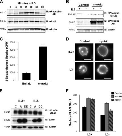
Figure 4.
Activated Akt regulates Glut1 trafficking to maintain surface Glut1 levels after IL3 withdrawal. (A) FL5.12 cells were withdrawn from IL3 for 6 h, restimulated with IL3 for various times, and immunoblotted for phospho-Akt, total Akt1 and actin. (B) Cells were transiently transfected with control or myrAkt vectors and cultured in the presence or absence of IL3 for 6 h. Phosphorylation levels of both mTOR and Akt were analyzed by immunoblot with actin as a loading control. (C) FL5.12 cells were transfected with either Bcl-xL or myrAkt expression vectors and cultured in the presence of IL3 for 18 h followed by a growth factor-withdrawal for 24 h and glucose uptake was measured. (D) Control or myrAkt-expressing cells were transfected with GFP-Glut1, and then they were cultured in the presence or absence of IL3 for 8 h, and GFP was visualized by fluorescence microscopy. Bar, 10 μm. (E and F) FLAG-Glut1 cells were transiently transfected with control vector (C), myrAkt (A), or a phospho-mimetic aspartate mutant of Akt (DD). Cells were cultured in the presence and absence of IL3 for 6 h. (E) Total FLAG-Glut1 and Akt1 levels were analyzed by immunoblot with actin as a loading control. (F) Surface levels of FLAG-Glut1 were measured by flow cytometry. Standard deviations of triplicate samples are shown. Representative results are shown for three or more repeated experiments.

