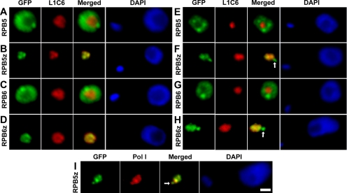
Figure 3.
Subnuclear localization of GFP-tagged RPB5 and RPB6 subunits. (A–D) Tsetse form procyclic cells fixed in 2% paraformaldehyde and labeled with anti-GFP and L1C6 anti-nucleolar antibodies. (E–H) Bloodstream-form cells processed in the same manner. (I) Bloodstream-form cells' colocalization of TbRBP5z-GFP with anti-Pol I largest subunit antibody. ESBs are indicated with white arrows. Bars, 1 μm.

